What are Varices?
The varices , also known as peripheral venous insufficiency , are 34 dilatations of the veins which, for various reasons, do not correctly comply with its function to bring the blood from return to the heart and, therefore, the blood accumulates in them, and dilate and return tortuous.
Usually the term varicose varicures are usually used to refer to those that appear on the legs, because it is the most frequent, but can also arise in other areas of the body as the esophagus (esophageal varices), the anal region (hemorrhoids) or In the testicles (varicocele).
The frequency with which they appear depends on many factors, but taking into account only those that give rise to clinical manifestations, it can be considered that between 10% and 15% of the population suffers from, increasing this percentage with age And sex, because there are more affected women than men.
Causes of varices
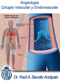
The veins are the vessels responsible for bringing the blood back to the heart, once they have irrigated all the tissues by bringing them oxygen and nutrients, which is called venous return .
We must bear in mind that, given the upright position of the human being, the blood of the legs must ascend, overcome the force of gravity, which means an added effort. To facilitate this task, the veins have in their interior valves that prevent the blood from receding, and also have the collaboration of the muscles of the legs that, when contracting, help to push the blood, establishing a unique sense towards the heart .
The planting pad also contributes to the correct development of this process. The pad is formed by a set of vessels that are filled with blood, like a sponge, and, by supporting the foot, the pressure exerted on the plant of it pushes that blood towards the heart.
When for some reason these valves can not fulfill their mission to prevent reflux, the blood accumulates, increasing the pressure, dilating and lengthening the veins (so they have to be written by forming nudos), and altering their wall, by What can come out liquid to the outside (extravasation) of the vein, altering the tissues of that area.
Varicose types
Varices Grade I or varicios
At this stage, they look at some sites and through the skin, the fine veins of violet color. Sometimes they can be starred shape, and vascular spiders are called. They are usually only an aesthetic problem but, at certain occasions, they can produce sensation of heaviness and fatigue on the legs.
Varices Grade II
The veins are becoming more visible and the first symptoms are noticed as:
- Heaviness and fatigue on the legs.
- Pain.
- Cramps.
- tingles.
- Heat feeling or chopping and scores.
Varices Grade III
The veins are more dilated and tortuous. Symptoms are progressively increasing, and swelling and edema and coloring changes appear on the skin.
Varices Grade IV
Eczematous areas and ulcers appear. Ulcers are difficult to treat and can be infected with ease.
Varicose diagnosis
The varicose diagnosis is very simple, and in many cases it is done by the patient itself. The exploration should be done standing, since this posture favors the appearance of varicose veins. At first glance, you see the dilated venous network, which indicates the situation and extension of the problem. In addition, you can also appreciate the coloration and appearance of the skin, the existence or not of other injuries such as spots, scraping lesions or ulcers, which allows assessing, in principle, the degree of affectation.
To the palpation the increase in venous tension and the existence or not of pain are observed.
With this data, a first evaluation of the importance of the problem is possible, which must be confirmed later with other tests as:
- Eco-Doppler: The most important test at the moment is the eco-Doppler, technique that combines ultrasound (to see the veins and arteries on its journey and check the alterations that may exist inIts interior) and the Doppler effect (in which most of the radars of traffic are based), which shows the venous flow and its anomalies.The test should be done with the patient standing and lying down.It is a non-painful test and that it does not need previous preparation.
- Flebography: Previously very used;It consists of injecting a iodized contrast into the vein and then performing an x-ray.It is almost discarded for being painful and present unnecessary risks, and its use is limited to very specific cases.
- Otras pruebas: hay más pruebas que pueden realizarse para el diagnóstico de las varices como: resonancia magnética (RNM), tomografía axial computerizada (TAC) yangiografía con isótopos. Pero, desde la aparición del eco-doppler apenas se utilizan.
Sclerotherapy
How is the procedure performed?
This procedure is done in the Office, with the use of a fine thin needle, the doctor injects the solvent solution from veins into the varicose veins and spider. The number of veins treated in a session varies, and depends on the size and site of the veins.
The procedure is normally completed in 30 A45 minutes.
How does scrolloopia work?
When the sclerosing solution is injected directly into the varicose veins or spider, irritates the vein layer, making it stick. With the passage of time, the glass becomes healing tissue that disappears from sight.
Will I need only one or more sessions?
Most patients will need several sclerotherapy sessions to completely treat their sick veins. In general, the largest veins are treated first and are allowed to heal to determine which of the small veins it needs to be treated. You must plan a minimum of 3 to 4sions to complete therapy. In addition, many patients will have veins that return over a period of time and these may require more sessions.
How long will I be out of work after the procedure?
After sclerotherapy, most patients can return to work on the same day. You must use the average compression or bandage according to the instructions for a minimum of 3 days. You should try to walk and avoid activities that prevent the movement of the legs such as bed rest or prolonged sitting. Usually, you will have a follow-up with your doctor from 7 to 14 days after the procedure.
